By Stuart Kerr, Technology Correspondent, LiveAIWire
A study published in Nature Cancer in 2026 found that Google’s mammography AI system detected 25 percent of the breast cancers that a standard screening programme would otherwise have missed as interval cancers, while matching the specificity of a human radiologist. Cancer detection rates in the study rose from 7.54 to 9.33 per 1,000 women screened. That is not a marginal improvement. For the women whose cancers were caught earlier rather than later, it may be the difference between early-stage treatment and a late-stage diagnosis.
The evidence base for AI in cancer diagnosis is accumulating faster than most patients or their doctors realise, and it is more complicated than the headlines suggest. The case for AI-assisted cancer screening is now substantial in specific domains, backed by genuine randomised trials as well as large feasibility studies. The case for autonomous AI diagnosis, algorithms replacing rather than supporting clinicians, remains contested, and the regulatory and ethical questions around it are nowhere close to resolved.
Table of Contents
Where the Evidence Is Actually Strongest
Breast cancer screening is the most rigorously tested application of AI in oncology, and it is worth being precise about which studies are which. The first genuine randomised controlled trial in this field, known as MASAI, involved more than 105,900 women in Sweden and found that AI-supported reading achieved higher sensitivity than standard double reading (80.5 percent versus 73.8 percent) with equivalent specificity, alongside a 12 percent reduction in aggressive or advanced cancers diagnosed in the following two years. An earlier interim safety analysis of the same trial, involving over 80,000 women, found AI-supported screening cut radiologists’ workload by 44 percent without increasing false positives.
Separately, Google’s mammography AI system was evaluated in a large two-phase study, a retrospective analysis of more than 115,000 mammograms followed by a prospective feasibility deployment across 12 sites. That is where the 25 percent interval-cancer detection figure and the 7.54 to 9.33 per 1,000 detection rate increase come from. It is important not to call this a randomised trial; it wasn’t one. But as a large-scale feasibility evaluation, it strengthens the same conclusion the MASAI trial reached through a different design.
A systematic review in BJR|Artificial Intelligence, the AI-focused sister journal of the British Journal of Radiology, found consistent evidence that AI assistance helps junior radiologists close the performance gap with more experienced clinicians, a genuinely useful finding for healthcare systems facing radiologist shortages.
What This Means If You’re the One Being Screened
For patients, the honest takeaway is that where AI is deployed properly and validated on a population similar to yours, the evidence now justifies its use in breast cancer screening specifically. It is a reasonable question to ask your radiologist or screening programme whether AI-assisted reading is part of their workflow, and increasingly, a programme not using it should be able to explain why. That said, the research base is far stronger for breast cancer than for most other cancer types, and the strength of the evidence should not be assumed to transfer automatically across cancers or care settings.
The Evidence Gap Nobody Likes Talking About
A meta-analysis of 49 randomised controlled trials, published in the Journal of the American College of Radiology and reported on by Diagnostic Imaging in mid-2026, found that colorectal cancer imaging accounted for nearly 80 percent of the trials reviewed. Pooled results showed AI use improved adenoma detection by 22 percent and polyp detection by 20 percent, though there was no significant improvement in detecting advanced adenomas or colorectal cancer itself. Single trials outside colorectal imaging suggested AI may improve breast cancer detection by around 20 percent, prostate cancer by around 40 percent, and high-risk oesophageal lesions by more than double.
The limitation the same meta-analysis identified is significant: none of the 49 trials it reviewed evaluated the impact of AI on actual patient outcomes such as survival. We know AI finds more abnormalities in several cancer types. We do not yet have the evidence to confirm that translates into better survival rates at population scale. That gap is not a reason for inaction, since earlier detection is a clinically reasonable proxy for better outcomes, but it is a reason for continued rigorous evaluation rather than unrestricted rollout.
Why the Research Doesn’t Match What Happens in Your Local Hospital
The practical deployment of AI in cancer diagnosis lags well behind the research literature. As of March 2026, the FDA had cleared 1,524 AI-enabled medical devices overall, with 1,163 of them, roughly 76 percent, in radiology specifically. But the distribution of those tools across healthcare systems is highly uneven. Major cancer centres with the resources to evaluate, implement, and maintain AI systems are adopting them steadily. Many community hospitals and screening programmes in under-resourced settings are not. The patients most likely to benefit from AI’s ability to catch subtle early-stage cancers, those without routine access to the most experienced radiologists, are also the least likely to be in systems currently using it.
There is also a bias problem that several 2025 reviews in the journal Cureus and elsewhere have flagged. AI systems trained predominantly on imaging data from specific populations can perform significantly worse on groups not well represented in that training data, and performance metrics reported in published research are often derived from cancer-enriched datasets that don’t reflect the proportion of cancerous findings in routine screening. When AI systems move from curated research datasets into real-world screening programmes, their performance frequently drops. Our own recent coverage of AI bias mitigation flagged an almost identical pattern in medical diagnosis broadly: a model can post 95 percent accuracy overall while performing at only 78 percent on a single demographic subgroup, a gap the aggregate number completely hides.
The Problem With an AI That Won’t Explain Itself
Most AI diagnostic tools currently in clinical use output a probability, cancer likely or not likely, without explaining which features of the image drove that conclusion. The BJR|Artificial Intelligence review flagged this as a real limitation on clinical adoption. Radiologists and oncologists are trained not just to identify findings but to justify their reasoning to patients, to colleagues, and in legal and regulatory contexts. A tool that says “70 percent probability of malignancy” without saying why is genuinely difficult to fit into that accountability structure, which is part of why our earlier coverage of explainable AI keeps surfacing across every high-stakes AI application, medicine included.
Regulators on both sides of the Atlantic are moving to require exactly this. The EU AI Act classifies medical diagnostic AI as high-risk, with core obligations for standalone high-risk systems applying from August 2026. AI embedded in devices already regulated under the EU’s Medical Device Regulation, which covers most diagnostic imaging AI, gets an extended deadline of August 2027, and the European Commission has proposed pushing that further to 2028 through a Digital Omnibus package still being negotiated as of this writing. Many of the FDA-cleared tools already on the market were approved before these explainability and bias-documentation standards existed, and bringing them into compliance retroactively is not straightforward.
Why Trust, Not Just Accuracy, Decides Whether Any of This Works
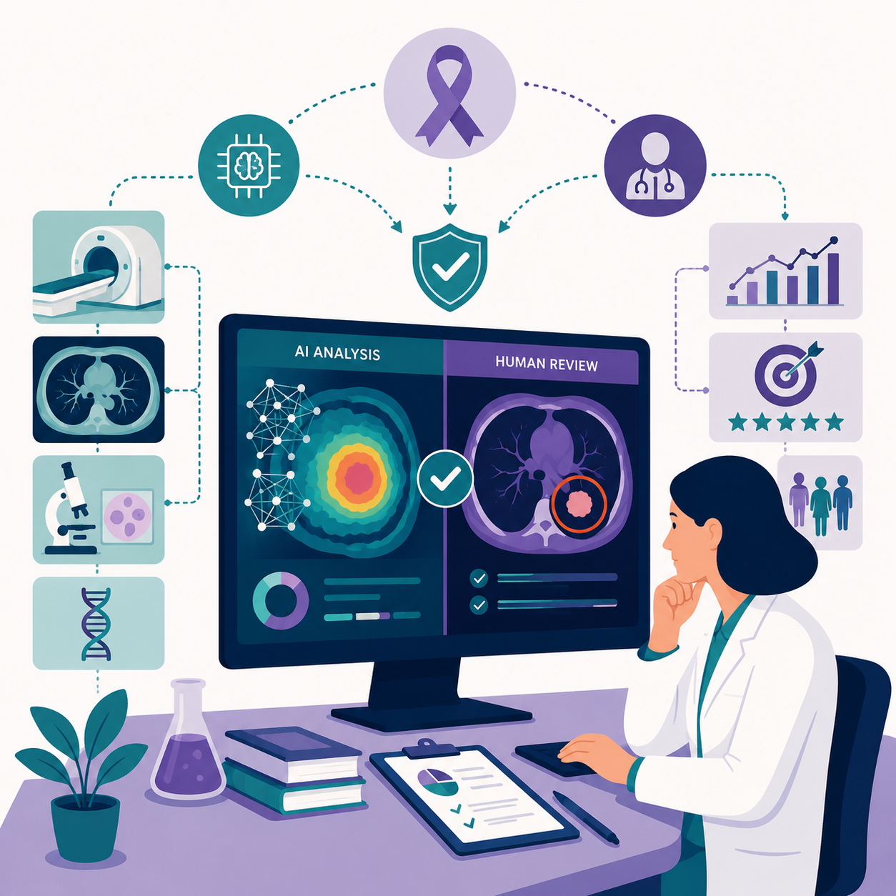
The most significant near-term challenge in AI cancer diagnosis isn’t technical capability. It’s workflow integration and clinician trust. Radiologist surveys consistently show that trust in AI tools depends heavily on how those tools are presented. Systems that show AI findings alongside the original image, letting the radiologist review the AI’s reasoning, generate far higher adoption than systems that present AI conclusions as final, unquestionable outputs. This mirrors a pattern we’ve seen repeatedly in AI deployment generally: the design of human-AI collaboration matters just as much as the underlying algorithm’s raw accuracy.
Early multimodal systems that combine imaging with a patient’s age, lab results, prior imaging history, and genetic information to generate risk-adjusted interpretations are now emerging in research settings. This is the direction the field is heading, and whether it will require entirely new regulatory frameworks, given that most existing clearance pathways were designed for single-input tools, is a live and unresolved policy question in both Washington and Brussels.
The Question Worth Asking Before Your Next Screening
For patients trying to understand where AI actually sits in their own cancer care, the honest answer depends enormously on which institution they attend, which cancer is being screened for, and whether the AI system in use has been validated on a population similar to their own. Asking directly whether AI-assisted reading is used, and whether it’s been checked for bias across different patient groups, is a reasonable and increasingly important question to bring to any appointment. The research supporting AI in breast cancer screening specifically is robust enough now that a lack of an answer should itself be a flag worth following up on.
Stuart Kerr is Technology Correspondent at LiveAIWire, covering artificial intelligence, emerging technology, and their impact on business, society, and everyday life. LiveAIWire publishes original AI journalism every weekday at liveaiwire.com.